In this article, we will delve into the fascinating world of chest X-rays and uncover the valuable information they provide about your health. A chest X-ray is a common diagnostic tool used by healthcare professionals to examine the structures within your chest, including the heart, lungs, and surrounding tissues. By analyzing the findings of a normal chest X-ray, medical experts can gain important insights into your overall well-being.
Understanding the Chest X-ray
The Basics
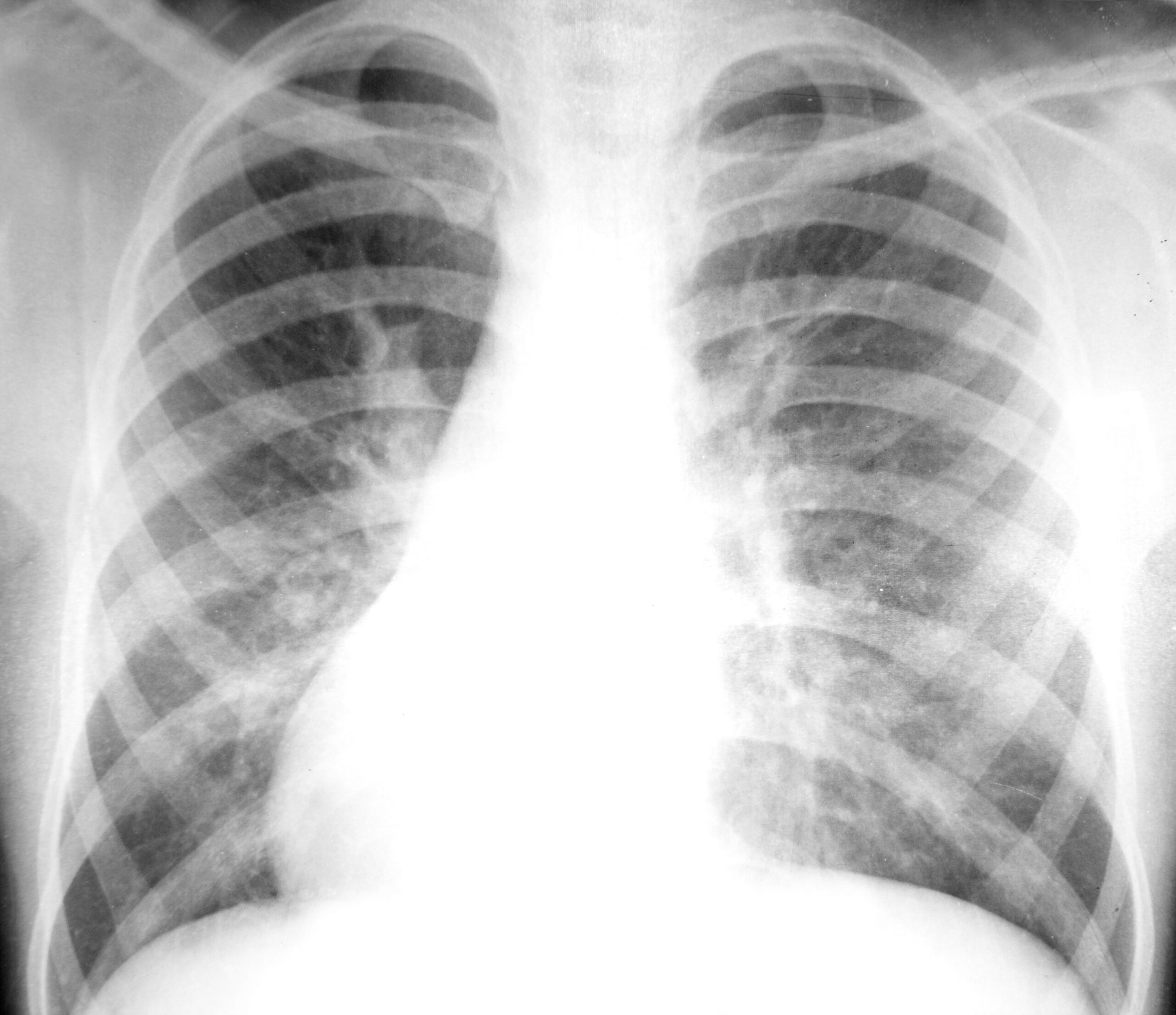
A chest X-ray, also known as a chest radiograph, is a non-invasive imaging technique that employs X-ray beams to generate detailed images of the chest’s internal structures. The X-ray machine emits a controlled amount of radiation that travels through your chest and produces an image on specialized film or a digital sensor.
Interpretation of the Results
When a radiologist assesses your health based on a chest X-ray, they meticulously consider a number of factors. These include the lungs, the heart, the bones, the blood vessels, and the adjacent tissues. By observing the size, shape, and position of these structures, they are able to identify any abnormalities or potential health issues.
Insights from a Normal Chest X-ray
Lungs and Airways
A standard chest X-ray can disclose vital respiratory information. It enables the radiologist to evaluate the condition of your lungs, detect any indications of infection or inflammation, and identify lung diseases like pneumonia, tuberculosis, and lung cancer. In addition, a chest X-ray can reveal the prevalence of chronic obstructive pulmonary disease (COPD) and asthma.
Cardiovascular System and Blood Vessels
A chest X-ray also evaluates the heart, which is an essential organ. It reveals the size, shape, and location of the heart’s chambers and valves. A normal chest X-ray can detect abnormalities of the heart, such as an enlarged heart or fluid accumulation around the heart. In addition, the radiologist is able to evaluate the condition of the blood vessels, identifying potential blockages or abnormalities.
Bones and Surrounding Tissues
Chest X-rays can provide valuable information about the chest’s bones and adjacent tissues. They are capable of detecting fractures, bone infections, and conditions such as osteoporosis. Additionally, thoracic X-rays can detect soft tissue abnormalities, such as tumors or masses.
Conclusion
A normal chest X-ray is a valuable tool for assessing your overall health and detecting potential abnormalities in your chest. By analyzing the lungs, heart, bones, and surrounding tissues, medical professionals can gain insights into various respiratory, cardiovascular, and musculoskeletal conditions.
In conclusion, a normal chest X-ray reveals important information about your health. It helps identify lung diseases, heart abnormalities, bone conditions, and other potential health concerns. Regular chest X-ray screenings, as recommended by your healthcare provider, can contribute to the early detection and timely treatment of various medical conditions.